Home
/ Knee Muscle Anatomy Mri - Pin by Balasubramanian on resonancia | Knee mri, Radiology ... - 1 november 2002 mri anatomy of the knee and shoulder james y.
Knee Muscle Anatomy Mri - Pin by Balasubramanian on resonancia | Knee mri, Radiology ... - 1 november 2002 mri anatomy of the knee and shoulder james y.
Knee Muscle Anatomy Mri - Pin by Balasubramanian on resonancia | Knee mri, Radiology ... - 1 november 2002 mri anatomy of the knee and shoulder james y.. Anatomy basic knee mri checklist. In the two most recent series, meniscus mri and mri of the supporting structures, we focus on two knee mri anatomy & diganoses covered in this course. Scroll through the structures to understand the anatomy. Knee, ankle, foot (2nd edition). Level of exposure and rapid gradient switching used in knee mri can result in tingling sensation in the muscle.
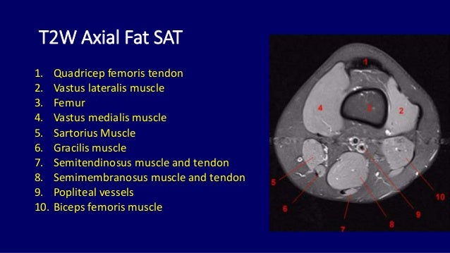
Scroll through the structures to understand the anatomy. This mri knee cross sectional anatomy tool is absolutely free to use. Musculoskeletal radiology south texas radiology group. 1 november 2002 mri anatomy of the knee and shoulder james y. 4, infrapatellar fat pad of hoffa.
MRI KNEE JOINT ANATOMY from image.slidesharecdn.com In the two most recent series, meniscus mri and mri of the supporting structures, we focus on two knee mri anatomy & diganoses covered in this course. Learn about mri anatomy with free interactive flashcards. Knee, ankle, foot (2nd edition). Free cross sectional anatomy of the knee based on mri : Tips to keep joints healthy. This long muscle flexes the knee. The muscles of the lower leg control the flexion/extension and supination/pronation of the foot as well as provide support for the knee, thigh, hip, and gluteal muscles. Normal mr imaging anatomy of the knee.
Mr arthrogram knee loose osteochondral lesion.
Normal mr imaging anatomy of the knee. Mri knee 1 by mohamed shaaban 6049 views. Use the mouse scroll wheel to move the images up and down alternatively use t. Scroll using the mouse wheel or the arrows. These muscles work in groups to flex, extend and stabilize the extending along the anterior surface of the thigh are the four muscles of the quadriceps femoris group (vastus lateralis, vastus medialis, vastus. Musculoskeletal radiology south texas radiology group. Involved early gray = muscle: Tips to keep joints healthy. Medical imaging technique used to examine the bones and soft tissue structures of the the mri has many advantages over other imaging techniques, one of them being its superior imaging anatomy: Scroll through the structures to understand the anatomy. Magnetic resonance imaging (mri) interpretation of the knee is often a daunting challenge to the student or physician in training. 1 november 2002 mri anatomy of the knee and shoulder james y. General anatomy and musculoskeletal system.
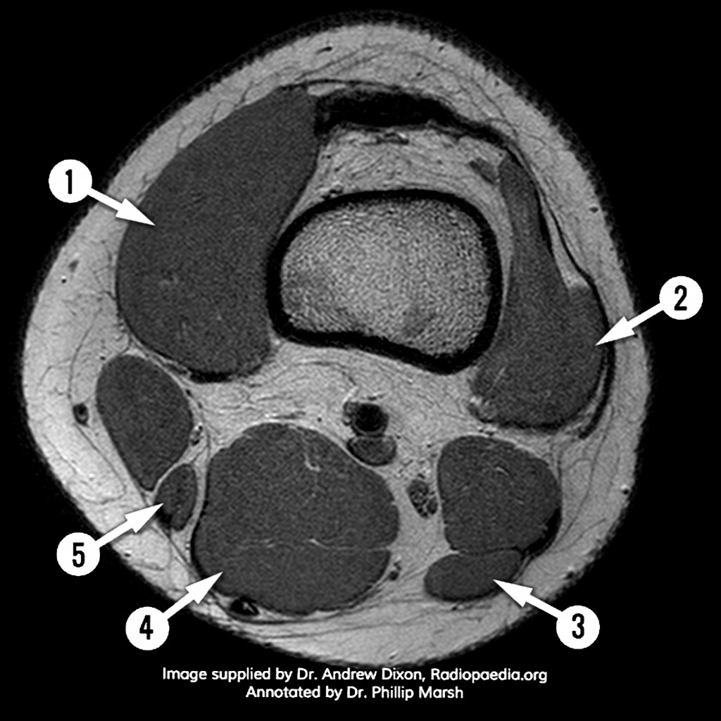
Level of exposure and rapid gradient switching used in knee mri can result in tingling sensation in the muscle. Anatomy basic knee mri checklist. This long muscle flexes the knee. Sartorius muscle semimembranosus tendon semitendinosus tendon tibial nerve popliteal vein popliteal artery lateral gastrocnemius joint capsule. Medical imaging technique used to examine the bones and soft tissue structures of the the mri has many advantages over other imaging techniques, one of them being its superior imaging anatomy:
Knee Muscle Anatomy Mri / Use The Mouse To Scroll Or The ... from prod-images-static.radiopaedia.org Properly performed and interpreted, mri not only contributes to diagnosis but also serves as an important guide to treatment planning and. Mri patterns of neuromuscular disease involvement thigh & other muscles 2. Tips to keep joints healthy. Learn about mri anatomy with free interactive flashcards. Medical imaging technique used to examine the bones and soft tissue structures of the the mri has many advantages over other imaging techniques, one of them being its superior imaging anatomy: Scroll through the structures to understand the anatomy. 1 november 2002 mri anatomy of the knee and shoulder james y. Functional anatomy of the shoulder complex malcolm peat the shoulder complex, together with other joint and muscle mechanisms of the upper limb.
Learn about the muscles, tendons, bones, and ligaments that comprise the knee joint anatomy.
Although not dangerous, can cause pain if exposure increases 50. Knee, ankle, foot (2nd edition). Magnetic resonance imaging (mri scan): Has stock or stock options held in conformis inc.; Serves as a paid consultant to or is an employee of conformis inc.; Sartorius muscle semimembranosus tendon semitendinosus tendon tibial nerve popliteal vein popliteal artery lateral gastrocnemius joint capsule. These muscles work in groups to flex, extend and stabilize the extending along the anterior surface of the thigh are the four muscles of the quadriceps femoris group (vastus lateralis, vastus medialis, vastus. Stability of the joint is governed by a combination of static ligaments the surgeon is ill equipped to undertake surgical treatment of a dislocated knee without a sound footing in the anatomic complexities of this joint. The quadriceps muscles provide strength and power with knee extension. Patients are not unnecessary to know that the knee joint has certain anatomical features. On anatomical parts the user. View of the anatomical labels. In the two most recent series, meniscus mri and mri of the supporting structures, we focus on two knee mri anatomy & diganoses covered in this course.
Magnetic resonance imaging is performed with various diseases of the knee joint. Involved early gray = muscle: Knee, ankle, foot (2nd edition). Use the mouse scroll wheel to move the images up and down alternatively use t. Use the checklist to quiz yourself.
Mri anatomy of knee Dr. Muhammad Bin Zulfiqar from image.slidesharecdn.com Click on the links to show each structure. General anatomy and musculoskeletal system. This webpage presents the anatomical structures found on knee mri. In the knee mri mastery courses, we give you everything you need in order to evaluate this joint. It is a complex mechanism that ensures the connection of the hip bone. Magnetic resonance imaging (mri scan): Involved early gray = muscle: Knee, ankle, foot (2nd edition).
Serves as a paid consultant to or is an employee of conformis inc.;
The knee joint is the junction of the thigh and leg. This mri knee cross sectional anatomy tool is absolutely free to use. View of the anatomical labels. Mr arthrogram knee loose osteochondral lesion. Song, uc san francisco msiv gillian lieberman md. Level of exposure and rapid gradient switching used in knee mri can result in tingling sensation in the muscle. Mri for evaluating knee pain in older patients: Medical imaging technique used to examine the bones and soft tissue structures of the the mri has many advantages over other imaging techniques, one of them being its superior imaging anatomy: The journal of musculoskeletal medicine. Although not dangerous, can cause pain if exposure increases 50. Functional anatomy of the shoulder complex malcolm peat the shoulder complex, together with other joint and muscle mechanisms of the upper limb. Free cross sectional anatomy of the knee based on mri : It is a complex mechanism that ensures the connection of the hip bone.